News
Improved treatments due to the influence of therapies on the epidermal cells
When performing any treatment in which the patient is moved, compressed, tractioned, rubbed or electrically or magnetically stimulated either with our hands or with machinery, we do not only act on the painful structures of the musculoskeletal system on which we intend to have an impact. Vascularization, fascia, nerves and viscera are also influenced.
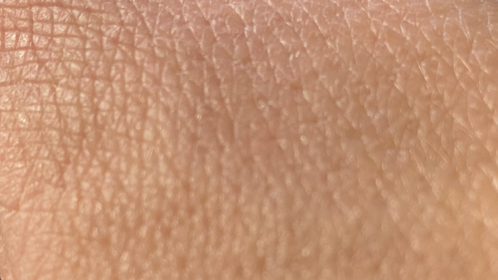
The first influence and effect when performing any treatment to the patient is on the epidermis, stimulating the Tactile Dome and Langerhans cells. Embryologically, they are of ectodermal origin as is the brain (Central Nervous System) and therefore send priority information from 0.04 mm to 1.6 mm. thickness of the epidermis.
Tactile domes are cellular complexes that are formed by clusters of1,2:
- Merkel cells specialized in tactile receptors of the epidermis.
- Keratinocytes.
- Dendrites of afferent sensory neurons.
The «Merkel discs or tactile dome» are sensory synapses and transmit tactile signals synaptically from Merkel cells to type A afferent nerve endings?1-3.
Light stimulation, simple, everyday light stimulation also elicits action potentials in A fibers? that innervate Merkel neuronal cell complexes. Since keratinocyte Cytokeratin 14 (CK14) is active in basal epidermal stem cells and governs channelrhodopsin-2 (ChR2) transcription, both keratinocytes and Merkel cells (Tactile Dome) can activate these nerve fibers since they are in close contact with the endings of the A-fibers. tactile1-3.
Merkel cells are specialized neuroendocrine cells of the epidermis. Keratinocytes play active roles as if they were the vanguard of an immune barrier, functioning as sentinels in response to skin damage or microbial intrusion. They are able to initiate and amplify a specific immune reaction through the activation of Langerhans cells that migrate to the lymph nodes draining the skin to recruit T cells to the skin, which secondly can be directly activated by keratinocytes2,3.
In addition, it is now well established that keratinocytes can also regulate noxious sensory neuronal activity. The identification of keratinocytes as primary transducers of noxious stimuli is a paradigm shift in the field of cutaneous sensory transduction4.
Current findings support the idea that keratinocytes, as activators of cutaneous neurons, play a central role in the initiation and maintenance of such abnormal transmission4.
Also in the epidermis are Langerhans cells, which are responsible for the uptake, processing and presentation of antigens to lymphocytes in the local lymph nodes. Their development, recruitment and retention in the epidermis is orchestrated by interactions with keratinocytes through multiple mechanisms5.
Therefore, simply touching a painful or dysfunctional area with our hands can improve the sensation or symptom due to the manual tactile activation and the reaction between the aberrant magnetic fields of the patient and the electromagnetic fields of the therapist, since living beings are a battery where action potentials and depolarizations of anions and cations are continuously being produced.
By producing an input when touching the patient, it acts on the epidermis sending direct information to the brain through the tactile dome (ectoderm) and on the dermis, creating force vectors and activating nerve receptors that influence the fascia, muscle and other metameric components (mesoderm). In addition, by the same action it also influences the local lymph nodes of the lymphatic system and therefore the whole organism.
Possibly these are the primary mechanisms and the explanation for the results obtained when performing inhibitory tests.
Magnetic Tape® acts on the epidermis as well as on tissues underlying the site of placement due to the magnetic properties it incorporates without the need to create tension with the tape. If tension is also created, the stimulation of the local tissues and the bandaged limb increases6.
Therefore, Magnetic Tape® is the only tool that acts on the Central Nervous System, on the Musculoskeletal System, on the Fascial System and on the Lymphatic System directly in a continuous, painless and non-invasive way.
Bibliography:
- Supp DM, Hahn JM, McFarland KL, Combs KA, Lloyd CM and Boyce ST. (2018). 372 Identification of Human Merkel Cells in Engineered Skin Substitutes Grafted to Mice. Journal of Burn Care & Research, 39 (suppl_1), S157-S157. DOI: http: //10.1093/jbcr/iry006.294
- Boulais N, Misery L. The epidermis: a sensory tissue. Eur J Dermatol. 2008; 18: 119-27. DOI: http://dx.doi.org/10.1684/ejd.2008.0348
- Jenkins BA, Foncetilla NM, Lu CP, Fuchs E, Lumpkin EA. The cellular basis of mechanosensory Merkel-cell innervation during development. eLife 2019; 8: e42633. DOI: 10.7554/eLife.42633
- Talagas M, Lebonvallet N, Berthod F, Misery L. Cutaneous nociception: Role of keratinocytes. Exp Dermatol. 2019; 28: 1466-1469. DOI: https://doi.org/10.1111/exd.13975
- Kaplan, D. Ontogeny and function of murine epidermal Langerhans cells. Nat Immunol 2017; 18:1068-1075. DOI: https://doi.org/10.1038/ni.3815
- Pamuk U and Yucesoy CA. MRI analyses show that kinesio taping affects much more than just the targeted superficial tissues and causes heterogeneous deformations within the whole limb. Journal of Biomechanics. 2015; 48 (16): 4262-4270. DOI: https://doi.org/10.1016/j.jbiomech.2015.10.036


